A karyotype test gives your doctor a complete picture of your chromosomes — the structures inside your cells that carry your DNA. If you or your baby might have a chromosomal condition like Down syndrome, this is often one of the first tests your doctor will order.
Here’s what karyotyping involves, who needs it, what it can and cannot detect, and what to expect before and after the test.
Key Takeaways
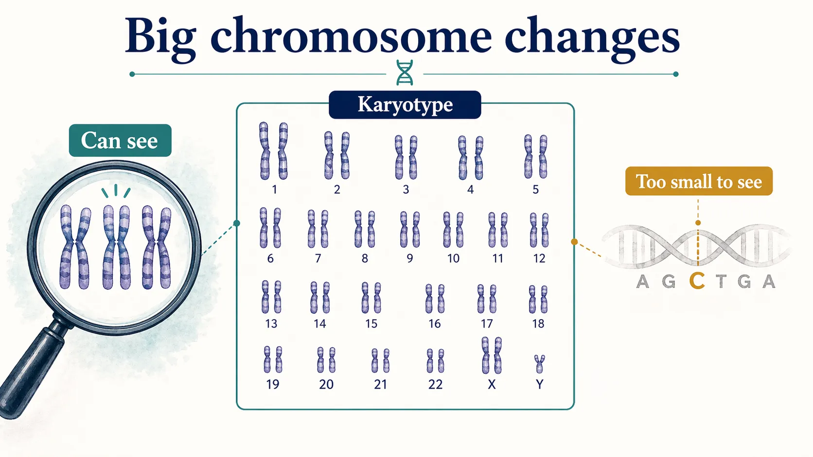
- A karyotype is a snapshot of your chromosomes - it shows whether you have the right number and whether their structure looks normal.
- Your doctor may order one to check for conditions like Down syndrome, Turner syndrome, or to investigate infertility or recurrent miscarriages.
- The test is affordable compared to more specialized genetic tests, and it catches chromosomal-level problems that other tests can miss.
- It has limits - karyotyping cannot detect small-scale genetic changes like those causing cystic fibrosis or sickle cell disease.
- Results require expert interpretation — always discuss your findings with a genetic counselor or your doctor.
How Karyotyping Works
During a karyotype test, a lab takes a sample of your cells and isolates the chromosomes. Technicians catch the chromosomes during cell division, specifically during the metaphase stage, when they are most visible, and arrange them in pairs from largest to smallest.
This organized image lets your doctor see whether any chromosomes are missing, duplicated, or structurally rearranged.
Why Choose Karyotyping Over Other Genetic Tests?
Karyotyping is more affordable than many specialized genetic tests, and it gives you a broad overview of all your chromosomes at once. It is especially good at detecting balanced rearrangements — structural changes in chromosomes that are often linked to infertility and that other tests can miss.
If your karyotype reveals something unusual, your doctor may order more targeted tests to confirm the findings or look at specific details more closely.
What Karyotyping Can Detect
Karyotyping is most useful for spotting chromosomal abnormalities — extra, missing, or rearranged chromosomes. Here are the most common conditions it can identify:
- Down Syndrome (Trisomy 21) - Caused by an extra chromosome 21. Affects physical features, cognitive development, and behavior.
- Edwards’ Syndrome (Trisomy 18) - An extra chromosome 18 leads to serious health problems including heart defects and low birth weight. Survival rates after birth are very low.
- Patau Syndrome (Trisomy 13) - An extra chromosome 13 causes severe heart problems and cognitive impairment. Most affected babies do not survive past the first year.
- Klinefelter Syndrome — An extra X chromosome in boys (XXY) that may delay puberty or affect fertility.
- Turner Syndrome and Mosaic Turner Syndrome — A missing or incomplete X chromosome in girls, which can affect the heart, height, and development.
What Karyotyping Cannot Detect
Despite its value, karyotyping has blind spots. It cannot reliably detect:
- Small-scale genetic changes - Mutations too small to see under a microscope
- Single-gene conditions - Disorders like cystic fibrosis, Tay-Sachs disease, sickle cell disease, and dwarfism caused by changes in individual genes rather than whole chromosomes
- Complex rearrangements - Chromosomal changes involving multiple chromosomes at once
- Marker chromosomes - Extra chromosome fragments that do not fit neatly into the standard 23 pairs
If your doctor suspects one of these conditions, they will likely recommend more specialized genetic testing alongside or instead of a karyotype. For a broader look at consumer-level options, see our guide to which DNA test is the most accurate.
How the Test is Done
The karyotyping process involves several lab steps. Here is what happens after your sample is collected:
- Cell culture - Your cells are placed in a nutrient-rich environment to encourage growth.
- Arresting cell division - A chemical called colchicine stops the cells at the metaphase stage, when chromosomes are easiest to see.
- Staining - A special dye binds to the DNA, creating banding patterns that help identify each chromosome.
- Imaging and arrangement - The lab photographs the stained chromosomes under a microscope and arranges them into a karyotype image by size and shape.
How Samples Are Collected
The type of sample depends on your situation. The most common options include:
- Blood draw — The simplest method. A standard blood sample provides white blood cells for analysis.
- Amniocentesis — If you are pregnant, your doctor may draw amniotic fluid using a thin needle inserted through your abdomen. This fluid contains fetal cells.
- Chorionic villus sampling (CVS) — Done between 10 and 13 weeks of pregnancy, CVS takes a small tissue sample from the placenta. Because placental tissue shares chromosomes with the fetus, it can reveal chromosomal conditions early. CVS is typically recommended for higher-risk pregnancies.
- Bone marrow — Used when your doctor suspects certain blood cancers or diseases.
Risks to Know About
A blood draw for karyotyping carries minimal risk — just the usual chance of minor bleeding or infection at the collection site.
Prenatal collection methods carry more risk. Here is a comparison:
| Amniotic Fluid Karyotyping Risks | Chorionic Villus Karyotyping Risks |
|---|---|
| Bleeding or leaking from the collection site | Miscarriage (very rare) |
| Some cramping or abdominal discomfort | Limb deficiencies if the test is done before 10 weeks |
| Infection (rare) | Abdominal discomfort and cramps |
| Preterm labor (rare) | Infection |
| Injury to the fetus from the needle touching them accidentally | Rupture in the amniotic sac |
The risks of amniotic fluid karyotyping increase if you are tested before 15 weeks of pregnancy.
Things to Consider Before Getting Tested
Before you decide on karyotyping, think about:
- Cost and insurance — Check whether your plan covers the test.
- Genetic counseling — A counselor can help you understand what the results might mean before you get tested.
- Procedure risks — Especially relevant if you are pregnant and considering amniocentesis or CVS.
- Emotional readiness — Some results can be difficult to process. It helps to have support in place.
What Happens After Your Karyotype
Follow your doctor’s aftercare instructions carefully. If you had an amniocentesis or CVS, you may need bed rest and should avoid driving for a short time.
Results typically take a few days to a couple of weeks. When they come back, do not try to interpret them on your own — your doctor or a genetic counselor can walk you through what the findings actually mean for you or your baby.
Being at increased risk for a chromosomal condition does not guarantee it will develop. Your doctor can help you understand the next steps and connect you with specialists if needed. If you want to learn more about your genetic health outside a clinical setting, our best health DNA tests roundup covers what consumer kits can and cannot tell you.